Imaging Modalities
Magnetic resonance imaging (MRI)


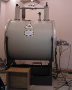
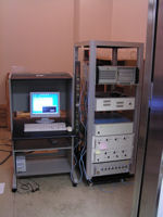
Kose and his coworkers developed a four-channel MR microscope equipped with a 2.34 T superconducting magnet[Matsuda et al., 2003]. Over 1,200 well-preserved human embryo specimens have been scanned in 128x128x256 image matrix. The voxel size varied from (40μm)3 to (150μm)3.
Episcopic fluorescence image capture (EFIC)


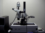
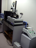
Episcopic fluorescence image capture (EFIC) is a 3D imaging method utilizing the tissue autofluorescence at the block face of the embedded sample. Using a sliding microtome, samples are sliced and the epifluorescence image on each surface is captured by a fluorescence microscope. The registered 2D image stacks are obtained, which may be used for rapid 3D rendering [Weninger, et al. 2002; Yamada, et al. 2010].
